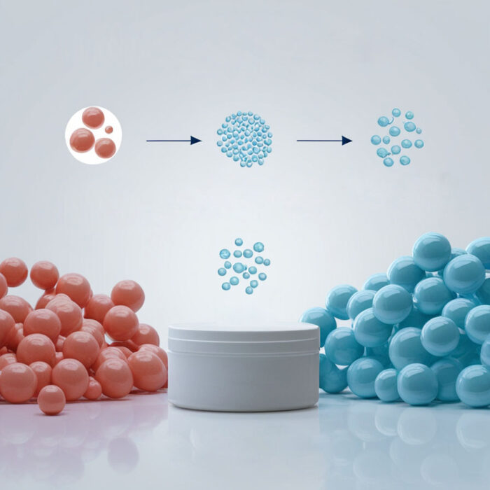
Ensuring that CD34+ cells are free from residual beads is more than a technical detail; it’s a matter of scientific integrity. Yet many labs underestimate how bead carryover can distort data and compromise reproducibility.
The Risks of Residual Beads
- Flow Cytometry Complications: Attached beads alter scatter properties, complicating gating and inflating CD34+ signals.
- Engraftment Distortion: Bead-bound cells may behave differently in vivo, slowing early kinetics.
- Quality and Compliance: While bead-labeled outputs are acceptable when disclosed for RUO applications, bead-free cells are preferred for in vivo work where standardization matters.
- Operational Inefficiencies: Detecting beads late forces labs to reprocess samples, adding cost and variability.
Microscopy as a QC Tool
Microscopy provides a reliable way to confirm bead absence. Clean CD34+ cells should appear uniform, free from refractile particles, and morphologically intact. By contrast, bead-labeled cells often display tethering, clustering, or irregular morphology.
FerroBio Advantage
FerroBio eliminates bead removal pitfalls at the source. Our platform delivers truly bead-free cells by design, requiring no additional washes or secondary bead-removal steps. Benefits include: – Clean cells validated by both flow cytometry and microscopy. – Stronger functional outcomes in NSG models. – Simplified workflows with fewer operator steps.
For labs scaling blood banking, research, or therapy preparation, FerroBio provides confidence that each vial contains what it should: clean, healthy CD34+ cells.
